
An injury to the face can be devastating to a person, often leaving unsightly scars that alter personal identity and self-confidence. At Dr. Amrika’s Aesthetics on Sarjapura Main Road, Bengaluru, the focus is centered on comprehensive scar minimization, immediate precision primary care, and advanced delayed scar revision. Whether managing an acute laceration within hours of trauma or re-sculpting a mature, weathered scar from years past, our clinical objective remains constant: restoring seamless structural harmony while delivering entirely subtle, natural-looking results that blend invisibly into your natural features.
Disclaimer: The medical information provided in this comprehensive guide is intended solely for educational purposes and patient reference. If you are experiencing an acute medical emergency, severe bleeding, or a compromised airway injury following trauma, please seek immediate emergency medical care at the nearest hospital.
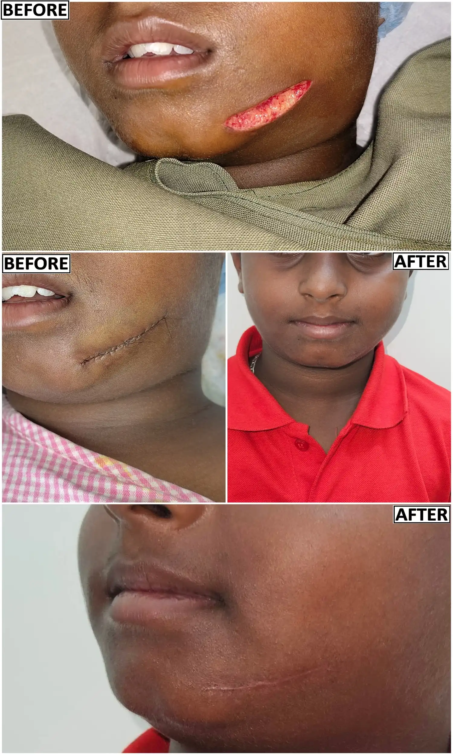
Mastering Facial Trauma & Scar Revision: Reconstructive Artistry, Fine-Suture Closure, and Advanced Scar Management in Pigmented Indian Skin Types
Section 1: Introduction to Facial Trauma and Reconstructive Artistry
The human face is our primary interface with the world. It is the canvas of our emotions, the cornerstone of our identity, and the most structurally complex and dynamic region of the human body. Unlike soft-tissue injuries to other parts of the integumentary system, a facial injury carries profound psychological, social, and functional implications. Because the face is constantly exposed to lighting, social interaction, and dynamic movement, any structural disruption, asymmetry, or poorly managed scar can fundamentally alter a person’s quality of life, self-esteem, and social relationships.
When an acute facial injury occurs—whether from a road traffic accident, a domestic mishap, a sports injury, or a dog bite—the immediate human instinct is to seek urgent medical care. However, the nature and surgical philosophy of that initial care dictates the long-term aesthetic outcome. A standard emergency room repair often prioritizes basic wound closure and rapid hemorrhage control over long-term cosmetic refinement. Standard sutures applied under tension across un-aligned dermal layers frequently leave permanent "railroad track" cross-hatch marks, depressed contours, or widened, rigid scars.
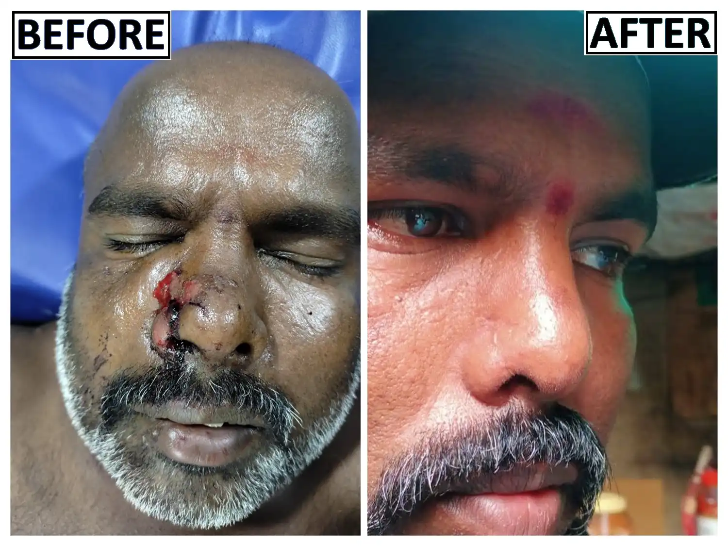
At Dr. Amrika’s Aesthetics, led by Dr. Amrika Seshadri, a dedicated Plastic and Reconstructive Surgeon with 11 years of clinical standing, we approach both acute facial injuries and mature scar deformities as complex exercises in reconstructive artistry. Our clinical philosophy centers on two dual pillars:
1. Multi-Layered Tension-Free Reconstruction: Utilizing specialized micro-surgical instruments, ultra-fine suture materials (6-0 and 7-0 monofilaments), and deep SMAS (Superficial Musculoaponeurotic System) load-bearing suspension.
2. Subtle, Natural Outcomes: Ensuring that every repair or revision honors the patient's underlying facial architecture without creating a pulled, tight, artificial, or overly surgical appearance.
Managing facial trauma and performing delayed scar revisions requires an advanced understanding of the distinct structural and cellular properties of thicker, more pigmented Indian skin types (Fitzpatrick Skin Phototypes IV, V, and VI). These skin types are biologically unique: while they possess a robust dermal architecture rich in collagen that resists environmental aging, they are exceptionally prone to pathological scarring, post-inflammatory hyperpigmentation (PIH), and keloid formation if subjected to rough tissue handling, harsh antiseptics, or improper tension.
Our clinic focuses entirely on the art and science of reconstructive salvage, scar revision, and aesthetic refinement. By employing precise anatomical realignment and a deep understanding of natural facial tension vectors, Dr. Amrika Seshadri ensures that both primary closures and delayed scar revisions minimize tissue distortion. Following any procedure, every patient is guided through a rigorous, multi-modality scar management protocol designed to suppress fibroblast overactivity, control melanocyte stimulation, and deliver the finest line scars possible.
Section 2: Comprehensive Facial Reconstruction: Acute Trauma vs. Delayed Injury Revision
While immediate primary care within the early hours of an injury prevents early distortion, millions of individuals live with old, mature, or disfiguring facial scars resulting from past emergency repairs, childhood falls, domestic accidents, or delayed healing. Dr. Amrika’s practice addresses both ends of the reconstructive spectrum with equal technical depth.
1. Acute Facial Trauma: The 72-Hour Golden Window
When a patient presents with a fresh facial laceration, puncture, or avulsion, the primary objective is to salvage every millimeter of viable tissue and execute a primary closure that eliminates surface tension entirely.
● Radical Conservatism: Because of the face's abundant blood supply, tissues that appear bruised, cyanotic, or partially detached often survive. We preserve all viable dermal margins.
● Debridement under Micro-Visualization: Removing microscopic dirt, debris, and devitalized cells using low-pressure saline irrigation rather than harsh scrubbing.
● Tension-Free Primary Alignment: Deep internal structural sutures absorb all physical mechanical stress, allowing the top epidermal layer to rest edge-to-edge without puckering.
2. Delayed Injury Revision & Old Scar Refreshing
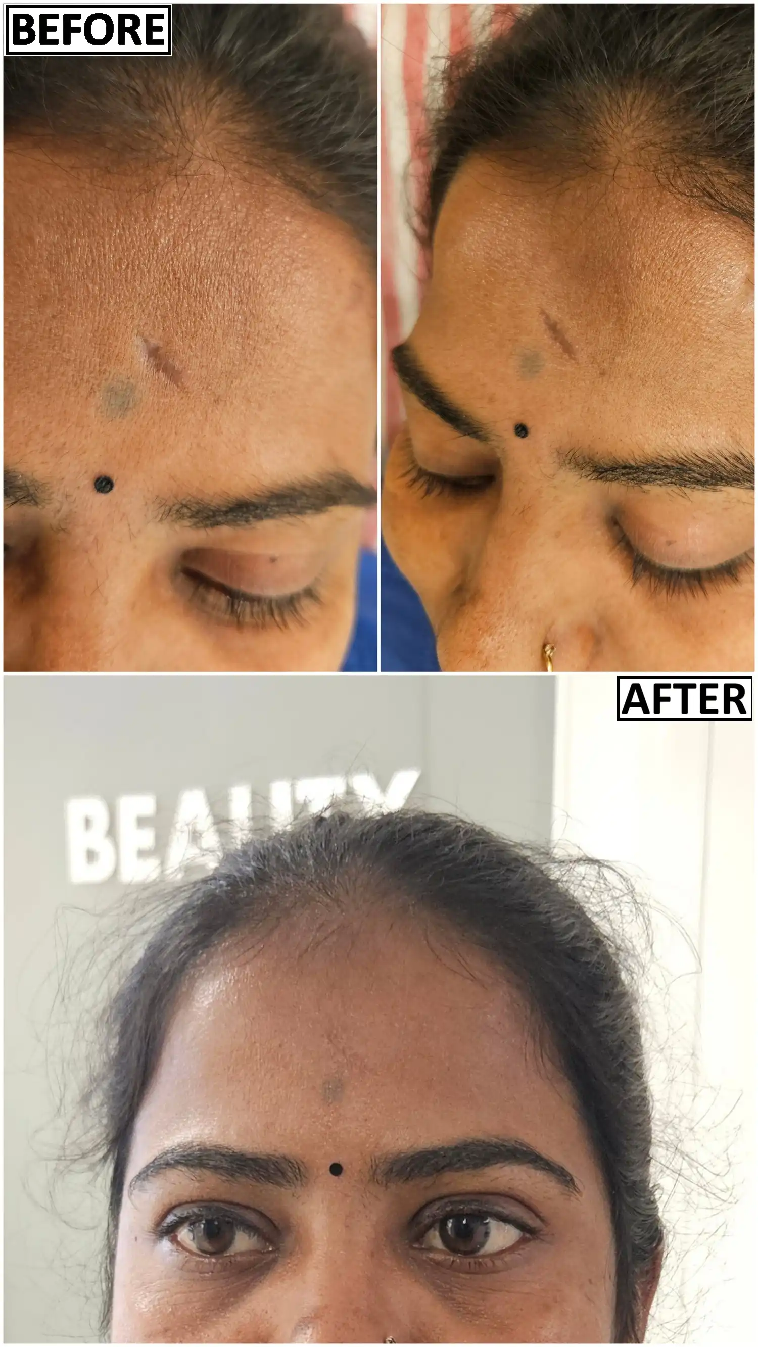
Old facial scars—whether wide, depressed, hyperpigmented, raised, or tethered to underlying facial muscles—do not have to be a lifelong burden. Dr. Amrika specializes in delayed scar revision, a meticulous process designed to remove, release, and refresh old scar tissue to recreate a clean, ideal healing environment.
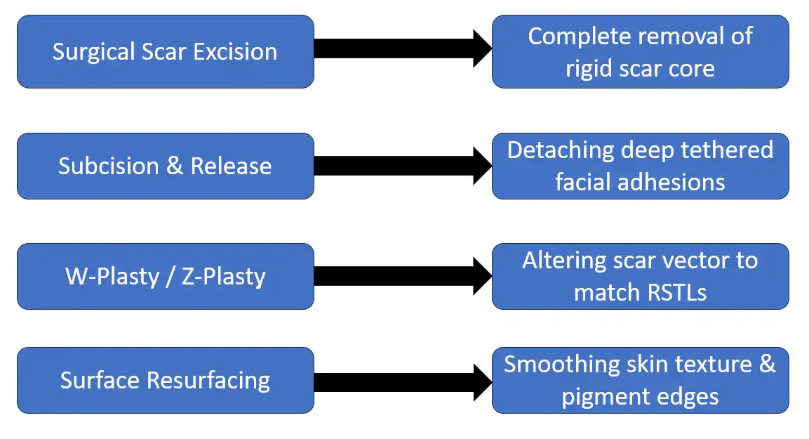
● Surgical Scar Excision & Re-Suturing: The old, disorganized scar tissue is surgically excised under microscopic precision, cutting back to healthy, pliable dermal margins. The fresh wound is then closed using our signature multi-layered, tension-free suture technique.
● Subcision and Dermal Release: Depressed or tethered scars often occur because fibrous bands pull the surface skin down toward the underlying facial fascia or muscle. Using specialized micro-blades, these fibrous tethers are gently released, allowing the skin to elevate back to its normal, natural plane.
● Geometric Vector Re-Direction (W-Plasty & Z-Plasty): Straight, rigid scars that run perpendicular to natural facial movement are constantly pulled apart by facial expressions. By converting a long straight scar into a series of small, interlocking zig-zags (W-plasty or Z-plasty), Dr. Amrika forces segments of the scar to lie parallel to natural expression lines, dramatically reducing mechanical pull and making the scar practically unnoticeable.
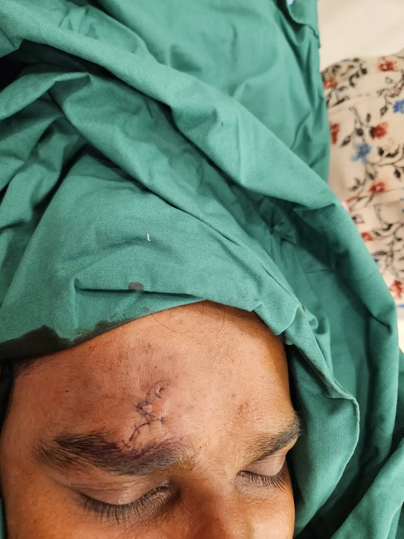
● Refreshing Weathered Scar Edges: For mature scars with irregular, stepped, or shiny surfaces, specialized surgical micro-dermabrasion or edge-blending techniques are used to feather the borders of the scar, blending it seamlessly into the surrounding healthy skin texture.
Section 3: Anatomical and Structural Pillars of the Face
To appreciate the complexity of managing facial lacerations, soft-tissue defects, and scar revisions, one must explore the specialized, multi-layered anatomy of the facial region. The face is not merely a sheet of skin draped over bone; it is a highly compartmentalized, three-dimensional system consisting of five distinct anatomical layers:
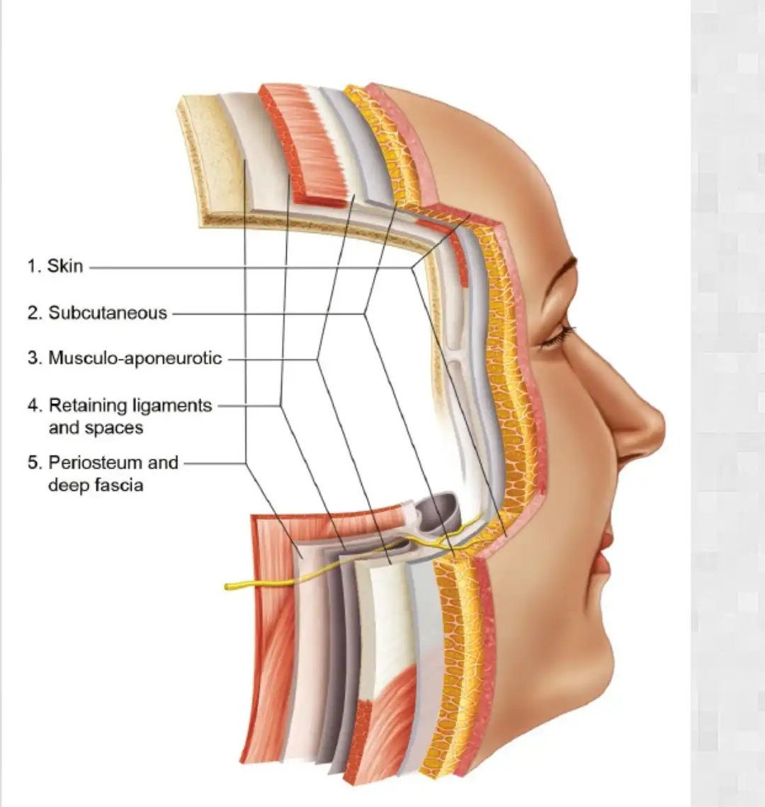
1. The Skin (Layer 1)
Facial skin varies dramatically in thickness, elasticity, and appendage density across different aesthetic zones. The skin of the upper and lower eyelids is among the thinnest in the human body, lacking a true subcutaneous fat layer, which makes it exceptionally unforgiving of poor suturing techniques or thick thread materials. Conversely, the skin of the nose, chin, and lower cheek is significantly thicker, densely populated with sebaceous glands, and deeply anchored to underlying structures. In Indian skin types, the dermis is structural denser and contains a higher concentration of active melanocytes and large, reactive fibroblasts. This biological profile means that any surgical trauma triggering a prolonged inflammatory cascade will inevitably result in post-inflammatory hyperpigmentation (PIH) or hypertrophic scarring if not managed delicately.
2. Subcutaneous Fat and Aesthetic Units (Layer 2)
Facial fat is partitioned into distinct superficial and deep compartments by fibrous septae. The arrangement of these compartments defines youthfulness, light reflection, and natural facial contours. When treating an acute laceration or revising an old scar, a plastic surgeon must strictly respect Facial Aesthetic Units—the forehead, brows, eyelids, nose, cheeks, lips, chin, and neck. If a laceration or old scar crosses the boundary between two aesthetic units (such as the vermilion border of the lip, the nasolabial fold, or the alar rim of the nose), even a slight misalignment creates an instantly noticeable asymmetry. Meticulous realignment of these topographical boundaries during closure or revision is essential for maintaining natural facial harmony.
3. The SMAS and Musculature Layer (Layer 3)
The Superficial Musculoaponeurotic System (SMAS) is a continuous fibromuscular network that invests the muscles of facial expression (mimetic muscles). Unlike skeletal muscles elsewhere in the body that insert into bones to move joints, facial muscles insert directly into the dermis of the skin. When a facial laceration or old scar penetrates deeply into the SMAS, the underlying muscles pull the wound edges apart every time the patient speaks, smiles, blinks, or frowns. If these dynamic physical forces are not neutralized during closure by placing deep, load-bearing suspension sutures into the SMAS layer, the resulting scar will inevitably stretch, widen, and become depressed over time.
4. Deep Loose Areolar Tissue (Layer 4)
This plane provides a sliding interface between the overlying musculoaponeurotic structures and the underlying bone. It is also the plane through which major neurovascular structures travel before sending terminal branches upward into the superficial layers.
5. Periosteum and Deep Fascia (Layer 5)
The deep structural framework anchored directly to the facial skeleton. In complex trauma involving deep facial cuts or old tethered scars, restoring continuity to this layer prevents long-term soft-tissue sagging or hollow contour defects.
Neurovascular Scaffolding
The face boasts a redundant, highly anastomotic blood supply derived from branches of both the internal and external carotid arteries (e.g., facial, superficial temporal, infraorbital, and ophthalmic arteries). While this rich vascularity leads to profuse bleeding during an acute injury, it is a massive advantage for the plastic surgeon. It allows for conservative debridement, as poorly perfused tissue fragments that would die in other body regions can often be salvaged on the face.
However, running alongside these blood vessels are critical neurological structures that demand expert surgical navigation during acute repair or deep scar revision:
● The Facial Nerve (CN VII): Governs all motor control of facial expression. It divides into five major branches (temporal, zygomatic, buccal, marginal mandibular, and cervical). The temporal branch (crossing the zygomatic arch) and the marginal mandibular branch (crossing the lower jawline) are superficial and exceptionally vulnerable to injury or accidental damage during deep scar releases.
● The Trigeminal Nerve (CN V): Provides precise sensory perception across the face via the ophthalmic (V1), maxillary (V2), and mandibular (V3) divisions. Injury to sensory nerve branches (like the supraorbital or infraorbital nerves) causes long-term numbness or painful neuropathic tingling if not meticulously identified and preserved.
Section 4: Pathophysiology of Indian Skin (Fitzpatrick Types IV–VI)

A successful primary facial repair or old scar revision cannot be executed without addressing the specific dermatological nuances of Indian skin types. The population across the Indian subcontinent predominantly falls within Fitzpatrick Skin Phototypes IV, V, and VI.
Understanding the cellular mechanics of pigmented skin explains why standard emergency room closures or aggressive surgical techniques often fail to produce optimal aesthetic results:
| Cellular Component | Biological Behavior | Clinical Impact |
|---|---|---|
| Melanocytes | Hyper-reactive, large melanosomes | High risk of PIH or hypopigmentation |
| Fibroblasts | Highly proliferative dense collagen production | Increased risk of hypertrophic scars |
| Sebaceous Glands | High density in T-zone and cheek areas | Risk of micro-incisions or suture reactivity |
| Baseline Skin Tension | Denser dermal matrix with firm elastic fibers | High outward pull on surface stitches |
1. Melanocyte Hyper-Reactivity & Pigmentary Instability
Pigmented skin contains larger, more melanized melanosomes that are distributed singly throughout keratinocytes rather than in clusters. When trauma occurs—or when surgical incisions are made during scar revision—inflammatory mediators (such as prostaglandins, leukotrienes, and histamines) stimulate these hyper-reactive melanocytes to rapidly increase melanin synthesis. This results in Post-Inflammatory Hyperpigmentation (PIH), causing the scar line or surrounding skin to turn a dark brown or purple hue.
Conversely, if the germinal layer of melanocytes is destroyed by rough tissue handling, high-tension pulling, aggressive chemical cleansers, or excessive electrocautery heat, permanent depigmentation (hypopigmentation) can occur. Hypopigmentation leaves a stark, chalk-white line that lacks melanin and is exceptionally difficult to camouflage. Preventing both PIH and hypopigmentation requires extreme surgical gentleness and immediate anti-inflammatory post-operative care.
2. Fibroblast Kinetics and Hypertrophic / Keloidal Tendencies
Histological studies of pigmented skin types demonstrate that dermal fibroblasts—the primary cells responsible for synthesizing collagen during wound healing—are larger, more numerous, and exhibit significantly higher proliferation rates compared to lighter skin types. When an acute injury heals under mechanical tension, or when a scar revision is performed without proper deep suspension, these hyper-active fibroblasts produce an overabundance of extracellular matrix components, specifically Type I and Type III collagen. This shift in cellular kinetics dramatically increases the risk of:
● Hypertrophic Scars: Raised, firm, red or dark lesions that remain confined within the original boundaries of the surgical incision or injury.
● Keloids: Aggressive fibroproliferative growths that extend beyond the margins of the initial injury, invading normal surrounding healthy tissue and causing itching, pain, and noticeable cosmetic distortion.
3. Increased Dermal Thickness and Sebaceous Density
The dermis in Indian skin is structurally thicker and contains a higher density of pilosebaceous units, particularly in the forehead, nose, and cheek regions. Sebaceous glands continuously secrete sebum, which can collect inside a poorly closed wound or revision site, creating a nidus for micro-infections or stitch granulomas. Furthermore, thicker skin possesses higher baseline tensile forces, meaning wound edges naturally pull apart with greater mechanical force than thinner skin types, placing immense physical demand on internal suture layers.
Section 5: Surgical Principles: Gentle Tissue Handling & Radical Conservatism
At Dr. Amrika’s Aesthetics, our operational approach to both acute facial trauma and delayed scar revision is grounded in two non-negotiable surgical dogmas: Gentle Tissue Handling and Radical Conservatism.
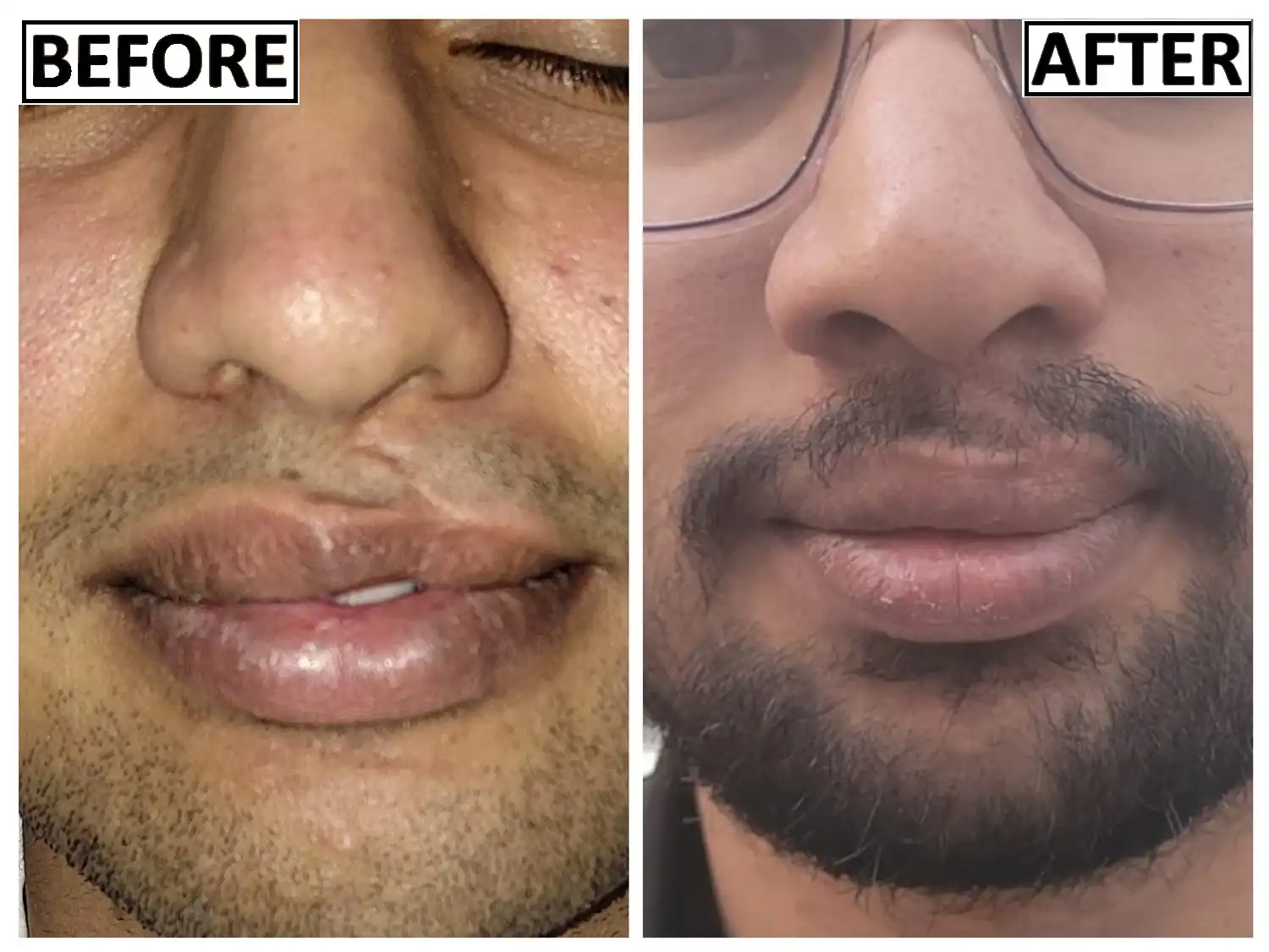
The Mechanics of Atraumatic Tissue Handling
Every touch of a surgical instrument inflicts cellular micro-trauma. When crushing forces or high-tension forceps are applied to skin margins, local capillaries are compressed, leading to focal tissue ischemia, cell necrosis, and a massive inflammatory cascade that directly fuels scar formation. Dr. Amrika Seshadri employs strict micro-surgical habits to protect delicate skin edges:
● Fine Skin Hooks Over Toothed Forceps: Standard toothed or smooth surgical forceps crush the dermal margins. We utilize ultra-fine skin hooks to gently retract and stabilize skin edges without compromising the delicate subdermal capillary plexus.
● Point-Specific Micro-Bipolar Electrocautery: While bleeding must be controlled, excessive use of standard electrocautery cooks surrounding dermal collagen, creating a wide zone of thermal necrosis that leads to wider scars. Bleeding is controlled using precise micro-bipolar coagulation or sustained, gentle pressure with warm saline-soaked gauze.
● Non-Traumatic Irrigation: In emergency settings, dirty wounds are often scrubbed vigorously with harsh chemical antiseptics like povidone-iodine or chlorhexidine. On facial skin, these agents destroy exposed, viable dermal cells. We utilize copious, low-pressure mechanical irrigation with sterile normal saline to flush away debris while preserving exposed cellular scaffolds.
Radical Conservatism in Debridement and Revision
In general surgical procedures, debridement often involves trimming wide margins of tissue until brisk bleeding is encountered. On the face, this approach can lead to catastrophic tissue loss, causing permanent distortion of eyes, lips, or nostrils. Because of the face's rich blood supply, tissues that appear bruised, swollen, or partially devitalized frequently recover if preserved carefully.
Dr. Amrika practices radical conservatism—removing only non-viable, necrotic tissue fragments and microscopic foreign debris, while preserving every possible millimeter of viable dermal edge during both acute repair and scar revision procedures.
Section 6: The Art of Tension-Free Fine-Suture Closure & Revision
Once an acute wound is thoroughly cleansed or an old scar is precisely excised, the reconstructive phase begins. The ultimate goal of plastic surgery closure is to re-approximate every anatomical layer perfectly, eliminate all internal dead space, and ensure that the final surface skin edges meet with zero mechanical tension.
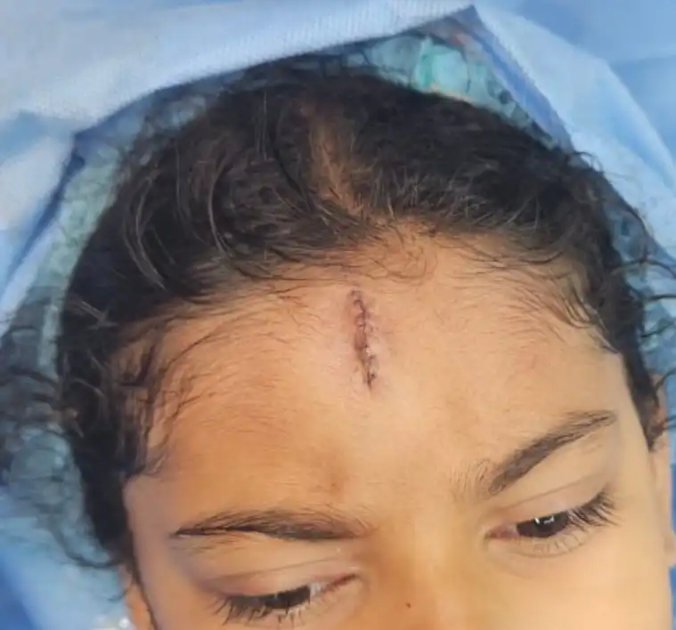
Multi-Layered Structural Realignment
Closing a deep facial cut or scar revision by simply running a stitch through the skin surface creates an underlying structural void (dead space) where blood and serous fluid collect. This predisposes the wound to micro-infections, delayed healing, and eventual scar depression. True long-term structural repair demands a multi-layered approach:
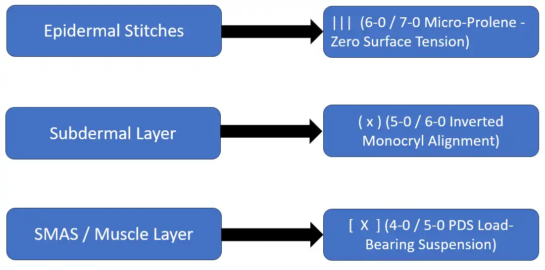
1. The Deep SMAS / Fascial Suspension Layer: The deep structural layer must bear the entire physical burden of facial movement and tension. We utilize long-lasting, absorbable monofilament sutures (such as 4-0 or 5-0 Polydioxanone/PDS) to re-approximate the SMAS and deep muscle fascia. These internal sutures act as an invisible structural suspension system, taking 100% of the mechanical tension off the outer skin surface.
2. The Subdermal Dermal Layer: To ensure the skin edges meet perfectly without stepping or overlapping, a dedicated layer of inverted, interrupted subcutaneous sutures (using 5-0 or 6-0 Monocryl or Vicryl) is placed. The knots are tied deeply so they do not push up against the surface epidermis. This step aligns the dermal-epidermal junction precisely.
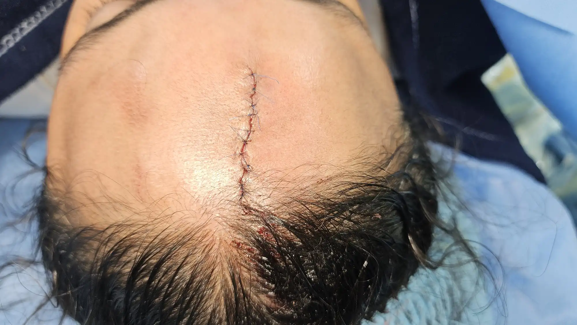
3. The Epidermal Layer (Fine-Suture Artistry): With the deep layers bearing all structural stress, the outermost skin layer requires only delicate positioning. We use ultra-fine, non-absorbable monofilaments (such as 6-0 or 7-0 Polypropylene/Prolene or Nylon) on micro-point precision needles. The stitches are placed microscopic distances from the wound edge and tied with just enough tension to approximate—never strangulate—the delicate skin margins.
Navigating Relaxed Skin Tension Lines (RSTLs)
The muscles of facial expression create continuous vectors of skin tension known as Relaxed Skin Tension Lines (RSTLs). These lines run perpendicular to the direction of underlying muscle contraction (for example, horizontal forehead lines run perpendicular to the vertical pull of the frontalis muscle).
● Parallel Injuries / Incisions: If a cut or surgical scar revision runs parallel to an RSTL, wound margins naturally fall together, yielding a thin, flat, practically invisible line.
● Perpendicular Injuries / Incisions: If a scar runs perpendicular to RSTLs, continuous muscle pulling constantly widens the healing tissue. In these cases, Dr. Amrika Seshadri utilizes local tissue rearrangements (such as W-plasty, Z-plasty, or geometric broken-line closures) to change the direction of the scar, forcing its segments to lie parallel to natural skin folds and blending the revision into natural expression shadows.
Section 7: Comprehensive Multi-Modality Scar Management Protocol
Whether performing a primary closure for acute trauma or executing a complex delayed scar revision, the surgical procedure is merely the first chapter in achieving an optimal outcome. The true battle against aggressive fibroblast activity and hyperpigmentation in Indian skin occurs during the remodeling phase of wound healing, which continues for 12 to 18 months post-treatment.
At Dr. Amrika’s Aesthetics, we enforce a mandatory, multi-modality scar management protocol tailored step-by-step to the patient’s healing trajectory:
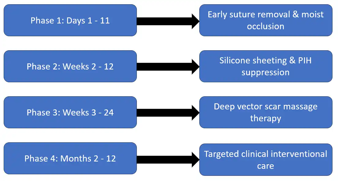
Phase 1: Immediate Post-Treatment Protection (Days 1 to 11)
● Early Suture Removal: Facial surface sutures are removed early—typically between day 7 and day 11 (and day 14 to 18 on the hands). Leaving skin sutures in longer allows epidermal cells to grow downward around the thread, creating permanent punctate "railroad track" marks.
● Moist Occlusive Healing: The wound or revision site is kept occluded with medical-grade ointments and hydration barriers to prevent dry scab formation. Hydrated skin epithelializes up to 50% faster than dry, crusted skin, reducing early inflammation.
● Strict UV Avoidance: Sunlight triggers hyper-reactive melanocytes, guaranteeing dark brown PIH in Indian skin. Strict sun avoidance and mechanical physical blocking (wide-brimmed hats, umbrellas) are enforced from day one.
Phase 2: Mechanical Stabilization and Occlusion (Weeks 2 to 12)
Once surface sutures are out and the epidermis is closed, the scar enters its most active collagen deposition phase.
● Medical-Grade Silicone Gel Sheeting: Silicone sheeting remains a gold standard in scar therapy. It works by creating a localized state of occlusion and deep hydration over the scar, which reduces capillary hyper-reactivity and signals fibroblasts to slow down chaotic collagen overproduction. Silicone sheets are worn for 12 to 24 hours daily.
● Topical Silicone Gels with Active Pigment Suppressors: For dynamic facial areas where sheets easily fall off, fast-drying medical silicone gel compounded with active lighteners (such as tranexamic acid, vitamin C, or azelaic acid) is used to simultaneously flatten the scar and suppress PIH formation.
Phase 3: Physical Disruption Therapy (Weeks 3 to 24)
● Deep Vector Scar Massage: Patients are taught a specific manual therapy technique. Using firm, circular friction perpendicular to the scar vector, patients massage the area for 3 minutes, twice daily. This physical pressure mechanically breaks up disorganized collagen tangles, forcing fibers to realign parallel to the skin surface. This dramatically reduces scar firmness, smooths surface irregularities, and prevents the scar from binding to underlying muscles.
Phase 4: Advanced Interventional Clinical Therapies (Months 2 to 12)
If a scar displays early signs of thickening, redness, persistent darkening, or uneven texture, we escalate to targeted clinical procedures:
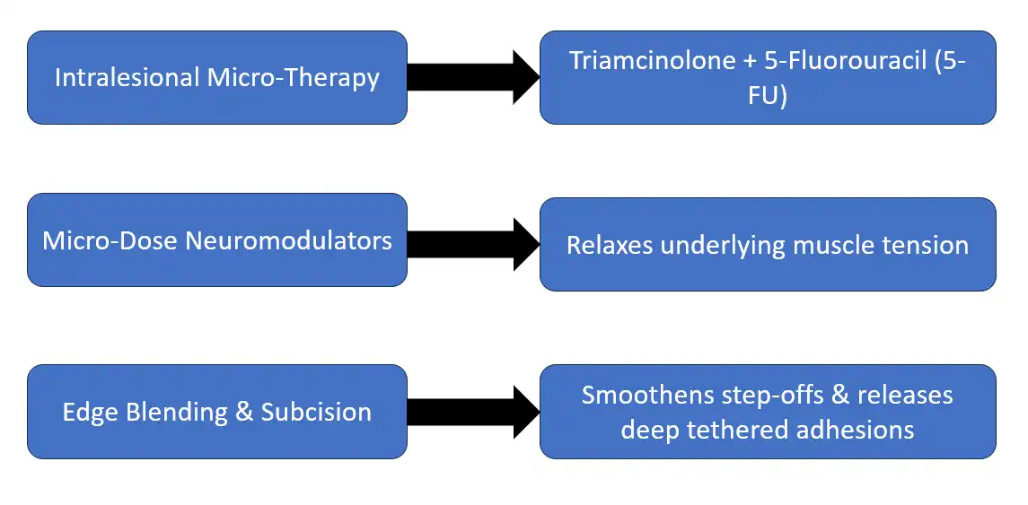
● Intralesional Micro-Therapy (Triamcinolone + 5-FU): For raised or itchy scars, a precise cocktail of Triamcinolone Acetonide (a corticosteroid) and 5-Fluorouracil (5-FU) is injected directly into the core of the scar. Corticosteroids halt inflammatory proliferation, while 5-FU stops excessive collagen synthesis. Combining both prevents the localized skin thinning (atrophy) or broken blood vessels (telangiectasia) often caused by steroids alone.
● Micro-Dose Neuromodulator Injections (Botox): Injecting micro-doses of botulinum toxin into the facial expression muscles directly beneath or surrounding a fresh or revised scar temporarily weakens those muscle fibers for 3 to 4 months. This stops continuous physical pulling on the healing dermal margins, acting as an invisible internal splint that allows the scar to mature thin and flat.
Section 8: Comprehensive Frequently Asked Questions (FAQ)
Timelines, Golden Windows & Scar Revisions
Q1: Why is it critical to seek expert treatment for a facial cut within the 72-hour golden window?
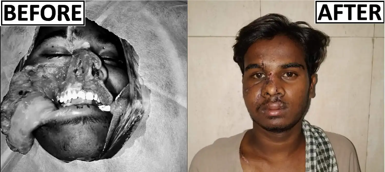
The first 72 hours following trauma represent the Golden Window for reconstructive success. During this timeframe, dermal edges remain viable, soft, and flexible, cellular viability at the margins is high, and the body's inflammatory response has not yet transitioned into a permanent matrix deposition phase. Furthermore, bacterial colonization spikes significantly after 24 to 48 hours. Closing a clean wound within 72 hours ensures minimal infection risk, zero surface tension, and the cleanest trajectory toward an invisible recovery.
Q2: What if I missed the 72-hour window, or have a facial scar that is several years old? Can Dr. Amrika still help me?
Yes, absolutely. Delayed scar revision is a major core specialty of Dr. Amrika Seshadri's practice. Whether a scar is 6 months old or 20 years old, mature scar tissue can be surgically excised, released from deep muscle tethers through subcision, or re-oriented using techniques like W-plasty. While acute treatment within 72 hours prevents initial bad scarring, delayed revision can completely transform an existing disfiguring scar into a fine, flat, and subtle line.
Q3: How does Dr. Amrika ensure that scar revision or trauma repair results look completely natural?
Natural, subtle outcomes are the cornerstone of our reconstructive philosophy. Over-tightened, pulled, or shiny scars occur when repairs are performed under high tension or without respecting natural facial contours. Dr. Amrika places structural suspension deep within the SMAS layer, allowing surface skin to rest naturally without tension. Additionally, scar lines are re-directed to sit directly inside natural expression folds and Relaxed Skin Tension Lines (RSTLs), ensuring the revised area moves naturally when you smile, speak, or express emotion.
Surgical Technique & Indian Skin Nuances
Q4: How does a specialist plastic surgery closure differ from a standard emergency room repair?
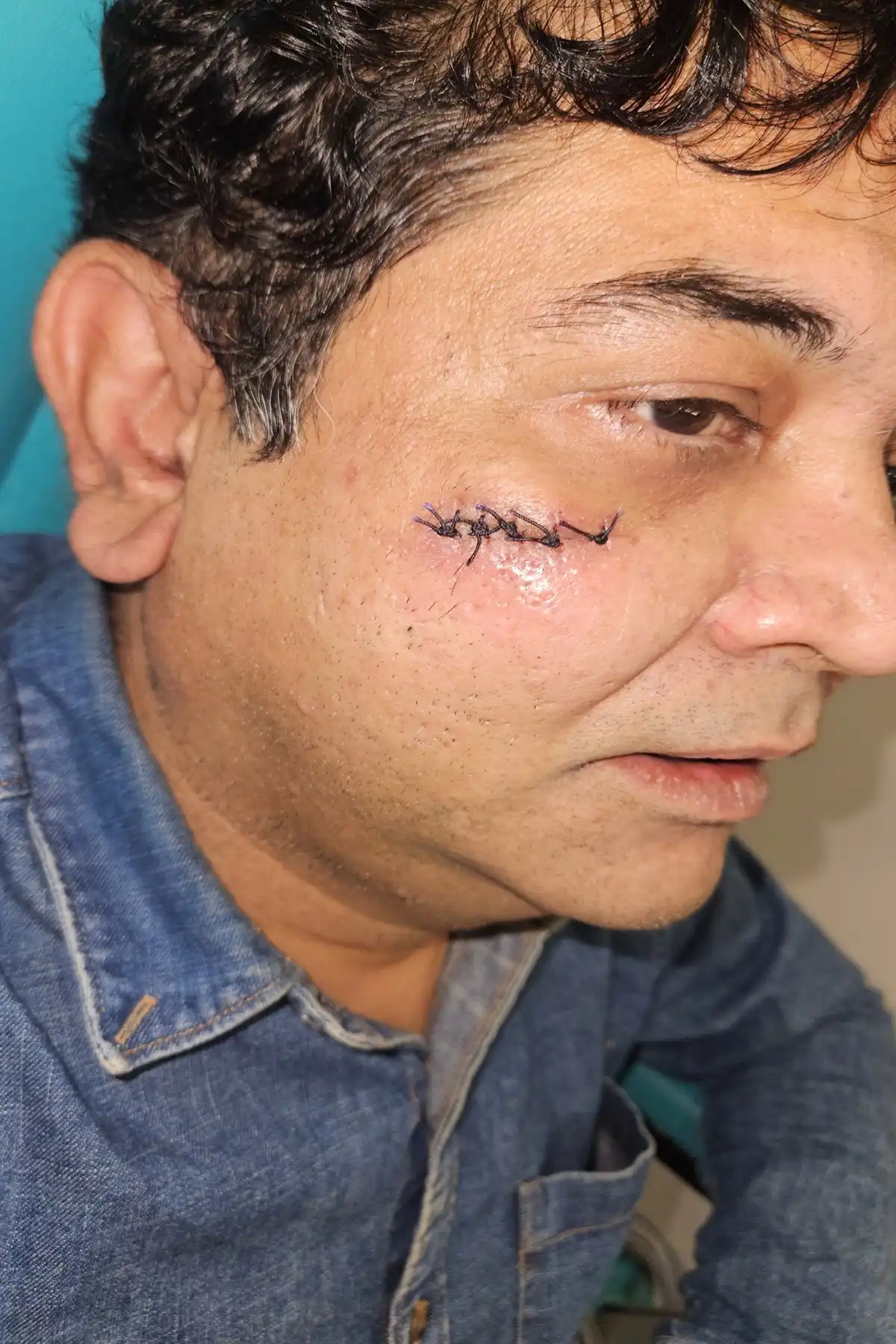
A standard emergency room repair focuses primarily on urgent hemorrhage control and rapid skin closure, often using thicker suture material placed through all skin layers simultaneously under high surface tension. While life-saving and necessary in trauma centers, this approach frequently leaves permanent "railroad track" cross-hatch marks and a wide, depressed scar. Dr. Amrika Seshadri approaches facial skin through a cosmetic reconstructive lens: utilizing deep structural suspension sutures to bear all mechanical stress, allowing the surface skin to be approximated using microscopic, ultra-fine sutures (size 6-0 or 7-0) under zero tension.
Q5: Why is Indian skin uniquely prone to dark or raised scars, and how is this managed?
Indian skin predominantly belongs to Fitzpatrick Skin Phototypes IV through VI, characterized by hyper-reactive melanocytes and highly active dermal fibroblasts. Any physical trauma, rough instrument handling, high tension, or infection triggers intense cellular reactions, leading to Post-Inflammatory Hyperpigmentation (PIH) or hypertrophic scar growth. Dr. Amrika manages this through strict non-traumatic tissue handling (using fine skin hooks instead of crushing forceps), non-thermal bleeding control, tension-free closures, and immediate post-operative pigment suppressors and silicone therapy.
Q6: Will facial trauma repair or scar revision require general anesthesia?
The vast majority of acute facial laceration repairs and elective delayed scar revisions are comfortably performed under targeted local nerve blocks or direct local anesthesia using ultra-fine needles. You remain entirely relaxed, awake, and completely pain-free throughout the precision repair, while avoiding the systemic risks, hospital stays, and prolonged recovery associated with general anesthesia. General anesthesia is reserved for very young children, highly anxious individuals, or extensive trauma involving underlying facial bone fractures.
Q7: What is a "W-plasty" or "Z-plasty," and when are they used in scar revision?
These are specialized local tissue rearrangement techniques. If a cut or old scar runs completely perpendicular to your natural facial lines, facial movements continuously pull the scar open, forcing it to stretch and widen. A W-plasty or Z-plasty breaks up a straight, tense scar line into a series of small, geometric, interlocking zig-zags. This alters the directional vector of the scar, forcing its segments to lie parallel to natural skin folds, which drastically reduces tension and blends the scar into natural facial shadows.
Post-Operative Care & Long-Term Results
Q8: Why must facial sutures be removed within 7 to 11 days, while sutures on arms or legs stay longer?
Facial skin possesses an exceptionally rich vascular supply, allowing dermal margins to adhere and heal significantly faster than skin on the limbs or trunk. If facial sutures are left in place past 11 days, surface epidermal cells grow downward along the suture track, creating permanent, indented punctate marks ("railroad tracking"). Removing outer stitches early (days 7 to 11) completely prevents these marks, while our deep internal SMAS sutures continue to hold structural layers together securely for months.
Q9: What immediate steps should I take at home if I sustain an acute facial injury?
1. Apply Continuous Direct Pressure: Use a clean, lint-free cloth or sterile gauze to hold continuous, steady pressure directly over the wound for 10 to 15 minutes to stop bleeding. Do not repeatedly lift the cloth to check.
2. Elevate Your Head: Sit upright or prop your head up on pillows to lower local blood pressure and reduce throbbing and swelling.
3. Rinse Gently with Saline or Water: Gently flush loose dirt or surface debris using sterile saline or clean running tap water. Do not scrub the wound, and avoid applying harsh rubbing alcohol, hydrogen peroxide, or thick home remedies, as these burn fragile cells and worsen long-term scarring.
4. Seek Specialist Plastic Surgery Care Immediately: Cover the area with a damp, clean dressing and contact Dr. Amrika’s Aesthetics or head to an emergency center to organize precision closure within the 72-hour golden window.
Q10: Can a deep facial scar that has turned dark brown or purple be restored?
Yes. Dark discoloration over a scar line is Post-Inflammatory Hyperpigmentation (PIH), caused by inflammation triggering excessive melanin synthesis in pigmented skin types. PIH is treated effectively through strict UV-blocking sunscreens, topical tyrosinase inhibitors (such as hydroquinone, kojic acid, tranexamic acid, and azelaic acid), and gentle clinical edge-blending therapies that normalize pigment distribution.
Q11: When can I begin wearing camouflage makeup over a repaired or revised facial scar?
You can safely apply makeup once the surface skin is completely closed and surface sutures have been removed for at least 3 to 5 days (typically around day 10 to 12 post-treatment). Always select hypoallergenic, non-comedogenic formulations, and apply makeup gently without rubbing the fresh scar tissue. Apply your topical silicone gel or sunscreen first, allowing it to dry fully before layering makeup over the area.
Q12: Is it possible to eliminate a deep facial scar so that skin is 100% restored to its original uninjured state?
Medical science cannot completely erase a deep dermal injury to return it to 100% untouched skin; any cut or surgical incision that penetrates into the deep dermis forms a permanent structural mark. However, by combining multi-layered internal suspension, tension-free micro-suturing within the 72-hour window (or precise delayed scar revision), and an assertive post-care protocol, Dr. Amrika Seshadri can refine even severe scars into thin, flat, soft lines that sit inside natural expression creases, making them practically unnoticeable to the naked eye.
Q13: Who is the most qualified doctor to treat facial cuts and scars, and is health insurance coverage applicable?
The most qualified specialist for facial trauma repair and scar revision is a Plastic and Reconstructive Surgeon, whose entire training is dedicated to delicate tissue handling, anatomical restoration, and minimal scarring. Dr. Amrika Seshadri brings 11 years of dedicated plastic surgery experience to every procedure.
Regarding financial coverage: Most health insurance policies cover acute traumatic facial injury repairs as medical necessities. At Dr. Amrika’s Aesthetics, a dedicated 100% insurance reimbursement support facility is available to assist patients with claim documentation and insurance processing.
Section 9: Long-Term Outlook and Reconstructive Philosophy
Recovering from a traumatic facial injury or living with a disfiguring old scar should never have to compromise your long-term confidence, social comfort, or identity. While an acute accident occurs in a split second—and old scars can feel like permanent reminders of past trauma—modern plastic surgery provides sophisticated solutions to restore facial aesthetics.
At Dr. Amrika’s Aesthetics, our clinical mission is to eliminate the long-term physical and social stigma of facial trauma. By blending deep structural stabilization with micro-suture cosmetic techniques and advanced delayed scar revision, Dr. Amrika Seshadri ensures that every layer of tissue is restored to its proper anatomical position. We do not merely close cuts or excise scars; we safeguard your unique facial expression, paying meticulous attention to the specific biological properties of Indian skin to deliver flat, soft, smooth, and naturally beautiful results.
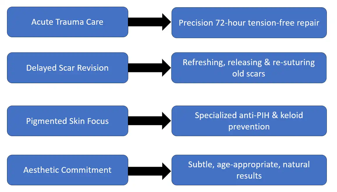
Secure an Expert Reconstructive or Scar Revision Evaluation
If you, a family member, or a patient has sustained an acute facial laceration, soft-tissue injury, or is seeking to refresh an old, disfiguring facial scar, do not compromise on specialized reconstructive care. Contact our clinical team directly to schedule a dedicated plastic surgery evaluation.
● Clinical Center: Dr. Amrika’s Aesthetics, Sarjapura Main Road, Bengaluru, India.
● Lead Specialist: Dr. Amrika Seshadri (Plastic and Reconstructive Surgeon, 11 Years of Standing).
● Consultation Protocols: Priority emergency scheduling is available for acute trauma repairs within the critical 72-hour window. Elective consultations are available for old scar revisions, subcision, and edge-blending evaluations.
Disclaimer: The medical information provided in this comprehensive guide is intended solely for educational purposes and patient reference. If you are experiencing an acute medical emergency, severe bleeding, or a compromised airway injury following trauma, please seek immediate emergency medical care at the nearest hospital.
Patient Reviews
Explore more
Related procedures
Get In Touch
Send a Message
Contact Information
Office Address
3rd floor SGR-2 Survey no 25/1A4
Above Style Union Sarjapura main road Doddakannelli
Bengaluru, Karnataka, 560035, India
Email: [email protected]
Phone: +91 9972951771
Website: www.dramrika.com
Social Media: